
1Julene B. Samuels, MD, FACS, Louisville, KY; 2The Female Pelvic Medicine Institute of Virginia, Richmond, VA
Presented at 38th American Society for Laser Medicine and Surgery Annual Conference on “Energy-based Medicine and Science”, April 11-15, 2018
Abstract
Background: Urinary incontinence (UI) has a prevalence of 30-40% in post-partum and menopausal women. Women may be reluctant to discuss UI with their healthcare providers as well as the degree to which it may negatively impact their quality of life (QoL) for numerous reasons including embarrassment and fear associated with treatment options. Women consistently express the preferred desire to address UI in a non-surgical and discreet manner. This study sought to report on results of a novel non-surgical treatment that may provide an affordable and discrete solution to this common problem.
Study Design/Materials and Method: This is a retrospective two-site study investigating the effectiveness of the treatment using quantified data, as well as the impact on QoL of incontinent women using a High-Intensity Focused Electromagnetic Technology (HIFEM) device. 20 women, 45 to 77 years (58.63±SD=9.86) who presented with urinary incontinence including stress, urge and mixed UI, were included in a pilot study. All patients completed a total of 6 treatments performed twice weekly for 3 consecutive weeks. Twenty patients completed King’s Health Questionnaire (KHQ) pre- and post-treatment. The same data was collected during 3 and 6-month follow-up as well. Additionally, patients reported the frequency of urinary leakage episodes and pad usage. Scores of the KHQ were calculated and statistically evaluated through t-test (p<0.05). The frequency of urinary leakage episodes and number of used hygienic pads were calculated through frequency of occurrence.
Results: Treatment with the HIFEM technology significantly improved QoL scores in all patients. There was a 60% improvement in both parts of the KHQ which were maintained through the 6-month follow-up (p<0.05). Nearly 75% of patients significantly decreased urinary leakage or achieved total dryness and maintained these results through follow-up. Pre-treatment, 16 patients used on average 2 hygienic pads per 24 hour period. During 3-month follow-up, 6 patients used 0.6 pads, 10 patients were completely dry. Twenty patients completed the 6-month follow-up, with eleven patients completely dry and 5 patients used 0.5 pads per 24 hour period. The vast majority of the patients decreased usage of hygienic pads to a minimum or totally eliminated usage.
Conclusion: Results suggest that HIFEM technology significantly improves the QoL and reduces UI in post-partum and menopausal female patients who present with all types of UI. This study confirms that further investigation is warranted.
1. INTRODUCTION
1.1. Prevalence of urinary incontinence
Urinary incontinence (UI) is defined as an involuntary loss of urine affecting mainly the female population. It is estimated that prevalence in young women is 20-30%, in mid-aged women 30-40%, whereas in elderly women prevalence rises to 50%.
1.2. Cause and consequence of urinary incontinence
The pelvic floor muscles (PFM) support pelvic organs and help control continence. Due to physiological changes such as body aging, childbirth or hormonal changes, PFM decondition and do not provide sufficient support for pelvic organs and continence control. This leads to PFM dysfunction with direct consequence toward incontinence.
1.3. Types of urinary incontinence and treatment options
There are 3 types of UI comprising stress urinary incontinence (SUI), urge incontinence and mixed urinary incontinence (MUI).
1.3.1. Stress urinary incontinence
Clinical symptoms of SUI are associated with involuntary urinary leakage during increased intra-abdominal pressure (e.g. coughing, sneezing, laughing, lifting etc.). The cause of the SUI is discoordination among weakened PFM and increased abdominal pressure. SUI is often associated with vaginal delivery, studies have shown 78.5 % of women were unable to contract pelvic floor muscles properly 1 year after delivery. SUI occurs as well in the post-menopausal period. Weakening of the pelvic floor muscles is caused by reduced estrogen level. In the case of SUI, treatment options range from PFM exercising (e.g. Kegel), intravaginal electrotherapy, hormone therapy, in addition to surgical intervention. Surgical intervention is recommended usually only in severe cases of SUI and a vast majority of female patients are reluctant to undergo surgical intervention, especially due to adverse events such as bleeding, development of urge incontinence due to inability to empty the bladder fully, and decreased sexual satisfaction.
1.3.2. Urge urinary incontinence
Urge incontinence is associated with an intense desire to void, during which the bladder pathologically contracts without cause. It is a neuromuscular dysfunction, typically representing a symptom of an underlying disease (e.g. diabetes mellitus). Traditional treatment of urge incontinence usually involves drug treatment.
1.3.3. Mixed urinary incontinence
A third type – mixed urinary incontinence (MUI) usually includes combination of stress and urge incontinence symptoms. MUI treatments usually involve a combination of PFM exercises and drug therapies. (1, 2)
1.4. Disadvantages of current treatment options
There are disadvantages to current treatment methods. In the case of SUI, one of the main problems in the case of pelvic floor exercising is the patients’ inability to selectively contract their pelvic floor muscles and to maintain an exercise routine. Kegel exercises are the most common form of PFM excercise, yet lack proof of efficiency as an effective solution. Another available treatment is intravaginal electrotherapy and biofeedback. A common concern of intravaginal electrostimulation is the electrode placements, which can cause patient’s discomfort. Adverse events can include bleeding, localized pain or irritation of the tissue under the patch electrodes. Surgical interventions are invasive and can also include adverse events. Pharmacotherapy is non-targeted, and side-effects such as dry mouth, bowel constipation and indigestion can occur. Today, both physicians and their patients seek a solution that meets the criteria of providing an effective clinical outcome via a non-invasive modality.
2. HIFEM technology
2.1. HIFEM technology Mechanism of Action
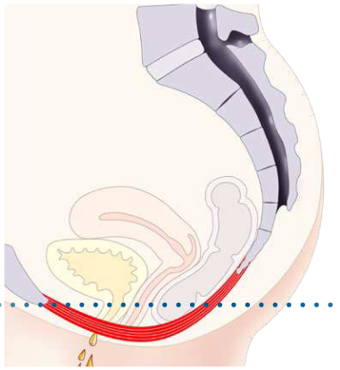
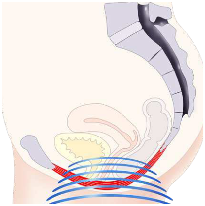
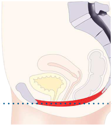
High-intensity Focused Electromagnetic technology (HIFEM) uses focused electromagnetic field with its intensity measured in Tesla. Such an intense electromagnetic field passes non-invasively through the pelvic floor area, interacts with PFM motoneurons and subsequently triggers supramaximal PFM contractions due to the action potential.
Text version of this figure
Three side-view drawings of the female pelvis, read from left to right and joined by arrows. First, the pelvic floor muscle (red) sags below a dotted reference line, with a downward arrow. Second, blue electromagnetic field lines reach the pelvic floor. Third, the muscle is lifted back up to the dotted line, with an upward arrow.
2.2. Supramaximal pelvic floor muscle contractions
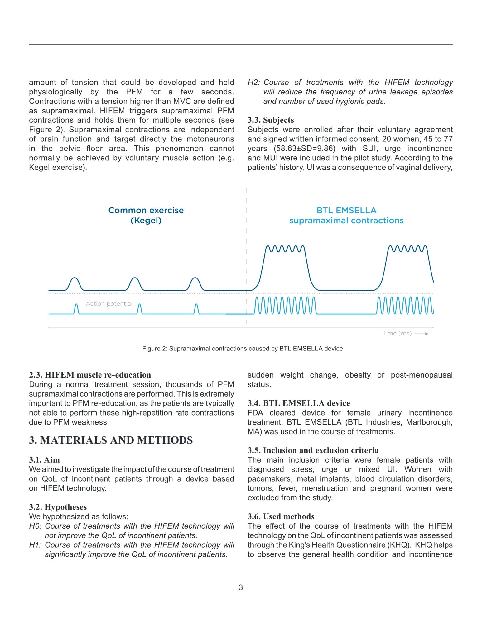
Maximal voluntary contraction (MVC) is the greatest amount of tension that could be developed and held physiologically by the PFM for a few seconds. Contractions with a tension higher than MVC are defined as supramaximal. HIFEM triggers supramaximal PFM contractions and holds them for multiple seconds (see Figure 2). Supramaximal contractions are independent of brain function and target directly the motoneurons in the pelvic floor area. This phenomenon cannot normally be achieved by voluntary muscle action (e.g. Kegel exercise).
Text version of this figure
Two traces plotted against time (ms), with a dashed line dividing the figure in two. Left, “Common exercise (Kegel)”: three short, low contractions, each with a single action potential. Right, “BTL EMSELLA supramaximal contractions”: two taller, longer contractions, each with a rapid series of action potentials.
2.3. HIFEM muscle re-education
During a normal treatment session, thousands of PFM supramaximal contractions are performed. This is extremely important to PFM re-education, as the patients are typically not able to perform these high-repetition rate contractions due to PFM weakness.
3. MATERIALS AND METHODS
3.1. Aim
We aimed to investigate the impact of the course of treatment on QoL of incontinent patients through a device based on HIFEM technology.
3.2. Hypotheses
We hypothesized as follows:
H0: Course of treatments with the HIFEM technology will not improve the QoL of incontinent patients.
H1: Course of treatments with the HIFEM technology will significantly improve the QoL of incontinent patients.
H2: Course of treatments with the HIFEM technology will reduce the frequency of urine leakage episodes and number of used hygienic pads.
3.3. Subjects
Subjects were enrolled after their voluntary agreement and signed written informed consent. 20 women, 45 to 77 years (58.63±SD=9.86) with SUI, urge incontinence and MUI were included in the pilot study. According to the patients’ history, UI was a consequence of vaginal delivery, sudden weight change, obesity or post-menopausal status.
3.4. BTL EMSELLA device
FDA cleared device for female urinary incontinence treatment. BTL EMSELLA (BTL Industries, Marlborough, MA) was used in the course of treatments.
3.5. Inclusion and exclusion criteria
The main inclusion criteria were female patients with diagnosed stress, urge or mixed UI. Women with pacemakers, metal implants, blood circulation disorders, tumors, fever, menstruation and pregnant women were excluded from the study.
3.6. Used methods
The effect of the course of treatments with the HIFEM technology on the QoL of incontinent patients was assessed through the King’s Health Questionnaire (KHQ). KHQ helps to observe the general health condition and incontinence impact on day-to-day life. Additional questions inquired regarding the number of used hygienic pads and frequency of urinary leakage.
4. DATA COLLECTION
4.1. Data collection
Data was collected pre- and post-treatment. The long-term effect was tested during 3- and 6-month follow-ups.
4.2. Therapy protocol
All patients completed 6 therapy sessions, 2 times per week. Patients were instructed by medical personnel to sit on the BTL EMSELLA chair with their spine straight, feet on the ground, hips, knees and ankles perpendicularly flexed. Throughout the procedure patients remained fully clothed. Therapy duration was set at 28 minutes; frequency range between 20-30 Hz with trapezoid intensity modulation were used to achieve gradual motor unit recruitment. Intensity (in %) was set according to patients’ feedback and comfort to achieve supramaximal PFM contractions.
4.3. Statistical evaluation
Data from the 20 patients were collected and statistically evaluated. During the course of treatment, no adverse events were recorded, and therapy was well-tolerated by all patients. KHQ scores were calculated (p<0.05). Results were compared between pre- and post-treatment, pre-treatment and 3- and 6-month follow-up data. Patients reported frequency of the urinary leakage episodes and use of hygienic pads, this data was then calculated as frequency of occurrence between pre- and post-treatment, as well as between pre-treatment and 3- and 6-month follow-ups.
5. RESULTS
5.1. The KHQ results
The results and hypotheses are discussed in the text below.
- H0: Course of treatments with the HIFEM technology will not improve the QoL of incontinent patients.
H0 hypothesis disproved. All patients (n=20) experienced improved QoL after course of treatment with the HIFEM technology, which was further proved by H1.
- H1: Course of treatments with the HIFEM technology will improve the QoL of incontinent patients.
H1 hypothesis proven.
5.1.1. KHQ Part 1 results
Pre-treatment average score of the KHQ-Part 1 was 92.22 points. Post-treatment average score of the KHQ-Part 1 decreased to 66.94 points. During 3-month follow-up, average score further decreased to 60.56 points, and to 37.04 points during 6-month follow-up, respectively. These scores are demonstrated as 50%, 51% and 60% levels of improvement in general health perception (p<0.05).
5.1.2. KHQ Part 2 results
Pre-treatment average score of the KHQ-Part 2 was 194.63 points. Post-treatment average score of the KHQ-Part 1 decreased to 154.44 points and was maintained during 3-month follow-up. During 6-month follow-up the score decreased to 90.59 points. These scores are demonstrated as 53%, 61% and 60% levels of improvement (p<0.05).
| Parameter | KHQ Part 1 | KHQ part 2 |
|---|---|---|
| Score pre-treatment (Mean±SD) | 92.22±36.09 | 194.63±107.34 |
| Score post-treatment (Mean±SD) | 66.94±34.91 | 154.44±104.23 |
| Score 3-month follow-up (Mean±SD) | 60.56±27.68 | 154.63±87.42 |
| Score 6-month follow-up (Mean±SD) | 37.04±34.44 | 90.59±90.79 |
| Level of improvement pre- and post-treatment (%) | 50% | 53% |
| Level of improvement pre-treatment and 3-month follow-up (%) | 51% | 61% |
| Level of improvement pre-treatment and 6-month follow-up (%) | 60% | 60% |
Legend: SD = standard deviation; KHQ = King’s Health Questionnaire
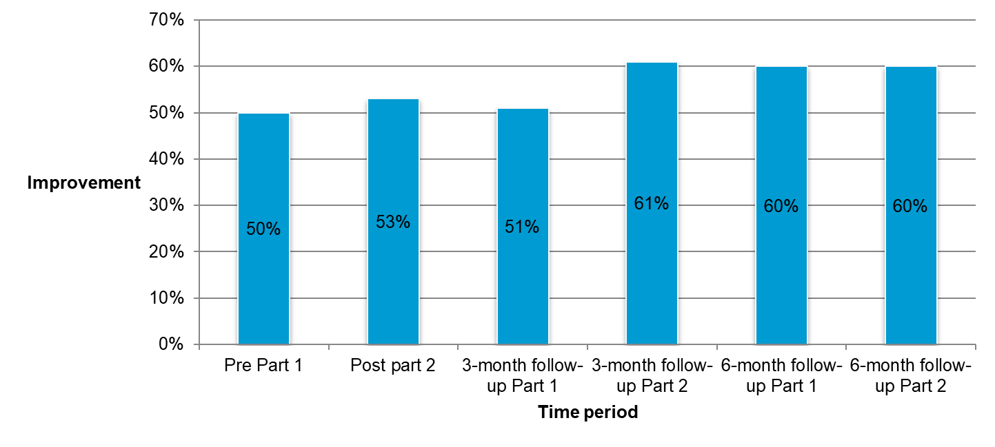
Text version of this figure
Bar chart. Vertical axis: Improvement, from 0% to 70%. Horizontal axis: Time period. Bars, left to right: Pre Part 1, 50%; Post part 2, 53%; 3-month follow-up Part 1, 51%; 3-month follow-up Part 2, 61%; 6-month follow-up Part 1, 60%; 6-month follow-up Part 2, 60%.
5.2. The urinary leakage episodes and use of hygienic pads
- H2: Course of treatments with the HIFEM technology will reduce the frequency of urine leakage episodes and number of used hygienic pads.
H2 hypothesis proven.
5.2.1. Urinary leakage episodes
Pre-treatment, all patients reported urine leakage in different severity (See Figure 6). Post-treatment, in 7 patients urine leakage episodes decreased to 1-3x a day, whereas 4 patients were completely dry. During 3-month follow-up, 7 patients decreased episodes to 1-3x a day, and another 11 patients to 1-3x a week, while 5 patients were completely dry. 20 patients completed the 6-month follow-up. 3 patients decreased episodes to 1x a day, whereas 12 decreased episodes to 1-3x a week, while 5 patients were completely dry.
| Frequency/number of patients | 5x a day | 3x a day | 2x a day | 1x a day | 3x a week | 2x a week | 1x a week | Never |
|---|---|---|---|---|---|---|---|---|
| Pre-treatment | 3 | 3 | 2 | 4 | 2 | 2 | 4 | 0 |
| Post-treatment | 2 | 2 | 2 | 3 | 3 | 3 | 1 | 4 |
| 3-month follow-up | 0 | 0 | 2 | 2 | 2 | 5 | 4 | 5 |
| 6-month follow-up | 0 | 0 | 0 | 3 | 2 | 4 | 6 | 5 |
5.2.2. Use of hygienic pads
Pre-treatment, 16 patients used on average 2 hygienic pads per 24 hour period. Post-treatment, 12 patients decreased use to 0.8 pad per 24 hour period, whereas 4 patients were completely dry. During 3-month follow-up, 6 patients were using 0.5-0.6 pad per 24 hour period, whereas 10 patients remained completely dry. At the 6-month follow-up, 5 patients were using 0.5-0.6 pad per 24 hour period, whereas 11 patients remained completely dry (See Figure 6).
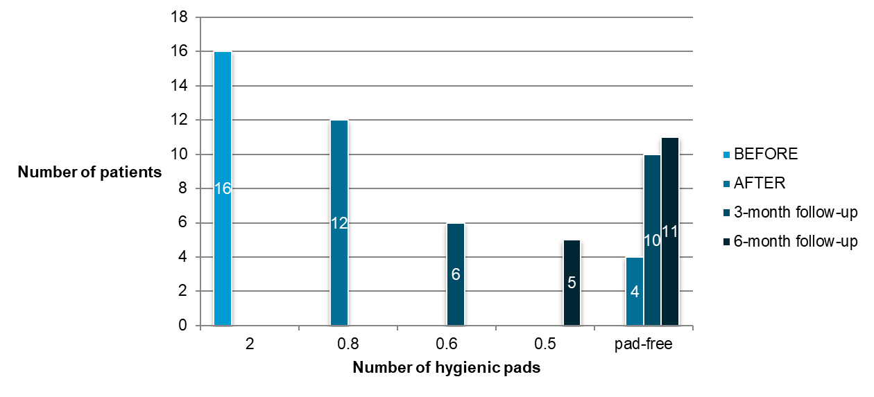
Text version of this figure
Bar chart. Vertical axis: Number of patients, from 0 to 18. Horizontal axis: Number of hygienic pads (2, 0.8, 0.6, 0.5, pad-free). Legend: BEFORE, AFTER, 3-month follow-up, 6-month follow-up. Bars: 16 patients before treatment at 2 pads; 12 patients after treatment at 0.8 pad; 6 patients at the 3-month follow-up at 0.6 pad; 5 patients at the 6-month follow-up at 0.5 pad; pad-free, 4 patients after treatment, 10 at the 3-month follow-up and 11 at the 6-month follow-up.
6. DISCUSSION
The results suggest that the treatment with HIFEM technology significantly decreases the negative impact incontinence has in patients’ day-to-day life. This improvement was observed in both short- and long-term results by KHQ, decreased frequency of urine leakage episodes, and decreased use of hygienic pads. The results are explained through myostimulation of the pelvic floor area by using high-intensity focused electromagnetic fields therapy, which trigger supramaximal PFM contractions. A single session brings thousands of PFM contractions. This is extremely important in PFM re-education helping the patients to regain PFM strength and bladder control.
7. CONCLUSION
UI represents an important healthcare problem with high prevalence and negative impact on patients’ QoL. As most of the patients are not suitable for current treatment methods, this study as well as previous research, suggest that UI can be treated non-invasively through HIFEM technology.
8. LIMITATIONS
The limitations of this study were the small number of patients and absence of a control group, such that a control randomized study with larger number of patients should take place in further research.
9. CONFLICT OF INTEREST
Authors declare that no conflict of interest exists.
10. REFERENCES
- Abrams P, Blaivas JG, Stanton SL, Andersen JT. The Standardisation of Terminology of Lower Urinary Tract Function. The International Continence Society Committee on Standartisation of Terminology. Scand d Suppl 1998; 114:5-19
- Abulhasan, J., Rumble, Y., Morgan, E., Slatter, W. and Grey, M. (2016). Peripheral Electrical and Magnetic Stimulation to Augment Resistance Training. Journal of Functional Morphology and Kinesiology, 1(3), pp.328-342
- Almeida FG, Bruschini H, Srougi M.: Urodynamic and clinical evaluation of 91 female patients with urinary incontinence treated with perineal magnetic stimulation: 1-year follow-up. J Urol. 2004 Apr; 171(4), pages 1571-4
- Bickford, R., Guidi, M., Fortesque, P. and Swenson, M. (1987). Magnetic stimulation of human peripheral nerve and brain. Neurosurgery, 20(1), pp.110-116.
- Bustamante, V., de Santa María, E., Gorostiza, A., Jiménez, U. and Gáldiz, J. (2010). Muscle training with repetitive magnetic stimulation of the quadriceps in severe COPD patients. Respiratory Medicine, 104(2), pp.237-245.
- Coletti, D., Teodori, L., Albertini, M., Rocchi, M., Pristerà, A., Fini, M., Molinaro, M. and Adamo, S. (2007). Static magnetic fields enhance skeletal muscle differentiation in vitro by improving myoblast alignment. Cytometry Part A, 71A(10), pp.846-856.
- Feldman M., Magnetic Stimulation for the Treatment of Urinary Incontinence in Women, California Technology Assessment Forum, San Francisco, CA, October 20, 2004
- Han T.R., Shin H.I., Kim I.S. Magnetic stimulation of the quadriceps femoris muscle: comparison of pain with electrical stimulation. Am J Phys Med Rehabil 2006; 85(7):593-599.
- Ishikawa N., Suda S., Sasaki T. et al., Development of a noninvasive treatment system for urinary incontinence using a functional continuous magnetic stimulator (FCMS) , Medical & Biological Engineering & Computing, 1998, 36, 704-710
- Man, W. (2004). Magnetic stimulation for the measurement of respiratory and skeletal muscle function. European Respiratory Journal, 24(5), pp.846-860.
- National Association for Incontinence (NAFC), www.nafc.org
- Ostrovidov, S., Hosseini, V., Ahadian, S., Fujie, T., Parthiban, S., Ramalingam, M., Bae, H., Kaji, H. and Khademhosseini, A. (2014). Skeletal Muscle Tissue Engineering: Methods to Form Skeletal Myotubes and Their Applications. Tissue Engineering Part B: Reviews, 20(5), pp.403-436.
- Sand PK, Richardson DA, Staskin DR. Pelvic floor electrical stimulation in the treatment of genuine stress incontinence: a multicenter, placebo-controlled trial. Am. J. Obstet. Gynecol. 1995; 173, pages 72–9
- StÖlting, M., Arnold, A., Haralampieva, D., Handschin, C., Sulser, T. and Eberli, D. (2016). Magnetic stimulation supports muscle and nerve regeneration after trauma in mice. Muscle & Nerve, 53(4), pp.598-607.
- Truijen G, Wyndaele JJ, Weyler J.: Conservative treatment of stress urinary incontinence in women: Who will benefit? Int Urogynecol J Pelvic Floor Dysfunct. 2001; 12(6), pages 386-90
- Wallis, M., Davies, E., Thalib, L. and Griffiths, S. (2011). Pelvic Static Magnetic Stimulation to Control Urinary Incontinence in Older Women: A Randomized Controlled Trial. Clinical Medicine & Research, 10(1), pp.7-14.
- Yamanishi T, Yasuda K, Suda S et al. Effect of functional continuous magnetic stimulation for urinary incontinence. J. Urol. 2000; 163, pages 456–9
- Yamanishi T, Yasuda K, Sakakibara R et al. Pelvic floor electrical stimulation in the treatment of stress incontinence: an investigational study and a placebo controlled double-blind trial. J. Urol. 1997; 158, pages 2127–31
- Yang, S., Jee, S., Hwang, S. and Sohn, M. (2017). Strengthening of Quadriceps by Neuromuscular Magnetic Stimulation in Healthy Subjects. PM&R.